Correction, 2026-09-08. This piece originally stated that whether the authors adjusted for kidney function was unconfirmed, and that the plasma NfL finding could therefore not be separated from renal confounding. That was wrong, and the answer was in the preprint all along: its main text reports that results were unaltered by further adjustment for estimated glomerular filtration rate. We found this only after extracting the full preprint PDF; the sentence appears once, closing the plasma-marker results section. The affected passages below have been corrected in place and marked. Two limits still stand: the adjusted estimate itself is never printed, and cystatin C is not mentioned anywhere in the paper — so this is the authors’ own reported robustness check, not an independent reproduction. The original caution was ours, not the paper’s, and withdrawing it makes this piece more favourable to the study, not less.
Evidence-first notes on bioscience and deep tech, at the edge of the lab and the market. Information only — not investment advice, and not medical advice. The paper discussed below is a medRxiv preprint (v1, posted 2026-08-12) that has not yet been peer-reviewed. It is an observational cohort analysis with Mendelian randomization as a secondary check, not a randomized controlled trial — nothing in it establishes causation, and nothing in this piece should be read as guidance to change anticoagulation therapy.
The 30-second version
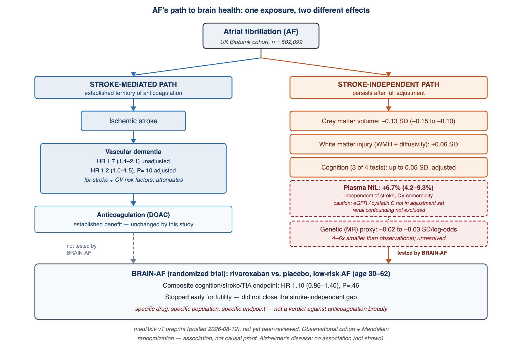
- What. A medRxiv preprint (v1, posted 2026-08-12, unpeer-reviewed) analyzes UK Biobank data on 502,099 people across five layers — brain MRI, cognitive testing, incident dementia, blood biomarkers, and genetics — to ask whether atrial fibrillation’s (AF) link to brain health runs through ischemic stroke or independently of it. After adjusting for stroke and cardiovascular risk factors, grey matter volume (−0.13 SD, 95% CI −0.15 to −0.10), white matter injury markers (+0.06 SD), cognitive test scores (small, up to 0.05 SD), and plasma neurofilament light chain (NfL, +6.7%, 4.2–9.3%) all persisted. Vascular dementia risk, by contrast, attenuated from HR 1.7 to HR 1.2 (P=.10) once stroke was accounted for — the authors read this as dementia being largely explained by ischaemic stroke, while brain-structure and cognitive changes are not.
- So what. This is an observational cohort study (plus Mendelian randomization as a triangulation check) — not a causal trial — and this preprint has not been peer-reviewed. Every effect size here is small: a population-average shift, not an individual diagnostic threshold. And the observational grey-matter estimate (−0.13 SD) is 4–6 times larger than the genetically-proxied one (−0.02 to −0.03 SD per log-odds of AF) — a gap the paper does not reconcile. Most importantly: the one biomarker carrying the “stroke-independent damage” story, plasma NfL, is cleared by the kidneys, and AF is strongly comorbid with chronic kidney disease — yet the reported fully-adjusted covariate set includes diabetes but not eGFR, creatinine, or cystatin C. [Corrected 2026-09-08] The preprint’s main text does state that results were unaltered by further adjustment for estimated glomerular filtration rate — one sentence, at the close of the plasma-marker results, which this outlet missed on first reading. The renal-confounding objection is answered by the authors’ own sensitivity analysis. Two limits remain: the adjusted estimate itself is never printed, and cystatin C appears nowhere in the paper.
- Now what. None of this is evidence to scale back anticoagulation — the data reconfirm that ischemic stroke, not some independent pathway, drives most of the clinical dementia risk, and anticoagulation’s established stroke-prevention benefit is untouched by this study. What has already been tested, and was negative, is narrower: in BRAIN-AF, a randomized trial of rivaroxaban versus placebo in low-risk AF patients aged 30–62, a composite cognitive-decline/stroke/TIA endpoint was not improved (HR 1.10, 95% CI 0.86–1.40, P=.46; stopped early for futility). That is a specific drug, a specific low-risk population, and a specific endpoint — not a general verdict against anticoagulation’s role in stroke prevention. And this preprint is not an Alzheimer’s disease story: the amyloid-adjacent marker GFAP showed no association (−0.01 NPX, P=.56) — this axis is vascular and axonal, not amyloid.
The five-minute read
Two tracks from one exposure
AF’s link to dementia is not new — the 2024 ESC atrial fibrillation guideline already cites adjusted associations with cognitive impairment (HR 1.39, 95% CI 1.25–1.53) and dementia (OR 1.6, 1.3–2.0), figures this outlet attributes to a secondary summary rather than the guideline text directly. What this preprint adds is a decomposition: the same 502,099-person cohort, split into five outcome layers, with stroke and cardiovascular risk factors adjusted out of each. Two different pictures emerge. The clinical diagnosis layer (vascular dementia) attenuates once stroke is added as a covariate — HR 1.7 (1.4–2.1) unadjusted falls to HR 1.2 (1.0–1.5, P=.10). The structural and molecular layers — grey matter volume, white matter hyperintensity, mean diffusivity, cognitive test scores, plasma NfL — do not attenuate; the effect sizes barely move after the same adjustment. The authors’ own words for the split, quoted directly and kept under 150 characters: “the association with dementia was largely explained by ischaemic stroke.” Brain structure and cognition were not.
Why the persisting layer needs a caveat before it becomes a story
The persisting layer’s only molecular marker is plasma NfL, a protein released by damaged axons — and also a protein cleared by the kidneys, with plasma levels rising as kidney function falls. Atrial fibrillation and chronic kidney disease are strongly comorbid in most cardiovascular cohorts. The preprint’s reported fully-adjusted covariate set (age, sex, deprivation, education, smoking, alcohol, BMI, blood pressure, lipids, diabetes, heart failure, coronary disease, hemorrhagic and ischemic stroke) includes diabetes but no direct kidney-function marker — no eGFR, no creatinine, no cystatin C. A companion astrocyte-injury marker, GFAP, showed no AF association at all (−0.01 NPX, P=.56, versus +0.18 NPX / 13.0% for actual ischemic stroke) — a pattern that is biologically coherent with axonal-not-astrocytic injury, but does nothing to rule out a kidney-function explanation for NfL specifically, since GFAP is cleared differently. [Corrected 2026-09-08] This has since been checked, and the answer was already in the preprint. Its own text reports that results were unaltered by further adjustment for estimated glomerular filtration rate; the sentence sits at the end of the plasma-marker results and applies to the protein markers, NfL among them. The association therefore survives the cheapest available falsification test, and the “stroke-independent axonal damage” reading keeps its molecular anchor. What is still missing is the adjusted figure itself, which the paper does not print, and any cystatin-C-based measure, which it never mentions — so this is the authors’ own reported robustness check, not an independent reproduction.
The trial that already tested this
The gap this preprint describes — a brain-structure and cognitive signal that does not track with stroke — is not purely hypothetical. BRAIN-AF already ran the most direct available test of whether anticoagulation closes it: rivaroxaban versus placebo in low-risk AF patients aged 30–62, composite endpoint of cognitive decline, stroke, and TIA. The trial found no benefit (HR 1.10, 95% CI 0.86–1.40, P=.46) and stopped early for futility; of 256 events, 234 (91.4%) were cognitive decline and only 13 (5.1%) were stroke. Read narrowly, that is the correct reading: a specific drug, in a specific low-risk younger population, on a specific composite endpoint, was negative. It is not a general statement about anticoagulation’s stroke-prevention value, which this preprint’s own dementia-layer finding reconfirms.
Deep dive
1. Background
The AF–dementia association is well established in outline. The 2024 ESC guideline for atrial fibrillation management cites adjusted cognitive impairment risk (HR 1.39, 95% CI 1.25–1.53) and dementia risk (OR 1.6, 1.3–2.0) — figures this outlet attributes to a secondary summary of the guideline, not a direct read of the guideline text. What has remained unresolved is whether that association is independent of AF’s cardiovascular comorbidities, in particular stroke — the preprint’s stated research question, quoted directly: it “remains unclear whether the association is independent of cardiovascular comorbidities, particularly stroke.” The authors are Alison Offer, Parag R. Gajendragadkar, Cornelia van Duijn, Paul M. Matthews, Barbara Casadei, and Jemma C. Hopewell, based at the University of Oxford’s Nuffield Department of Population Health, Imperial College London, and the UK Dementia Research Institute.
2. What this study newly shows
The paper’s contribution is not a new AF–brain association — it is a same-cohort decomposition across five outcome layers that shows the stroke contribution differs by layer. Imaging: full-cohort grey matter volume falls −0.13 SD (−0.15 to −0.10) after full adjustment, with regional loss across frontal, parietal, temporal, occipital, and cerebellar cortex plus hippocampus and thalamus — the amygdala was the one region showing no association. White matter hyperintensity burden and mean diffusivity each rise 0.06 SD (0.03–0.10). Cognition: after adjustment, 3 of 4 tests retained a small but statistically significant effect (up to 0.05 SD, P<.01). Dementia: vascular dementia risk fell from HR 1.7 (1.4–2.1) to HR 1.2 (1.0–1.5, P=.10) after adding stroke and cardiovascular covariates; Alzheimer’s disease showed no association at any stage. Biomarkers: plasma NfL rose 0.09 NPX (6.7%, 4.2–9.3%, P<.0001), independent of stroke and cardiovascular comorbidity in the reported model, while plasma GFAP showed no association (−0.01 NPX, P=.56) — for comparison, actual ischemic stroke raised GFAP by 0.18 NPX (13.0%). Genetics: a 269-variant Mendelian randomization instrument for AF (from Roselli et al., cited within the preprint, not independently accessed by this outlet) showed the same directional pattern — vascular dementia OR 1.21 (1.10–1.34) attenuating to 1.09 (0.96–1.25, P=.18) after adjusting for the instrument’s ischemic stroke effect (via GIGASTROKE consortium summary statistics, also cited within the preprint), while the grey-matter genetic estimate held at −0.02 to −0.03 SD per log-odds of AF liability.
The GFAP-null/NfL-positive contrast gives the persisting layer some internal biological coherence: GFAP marks astrocyte reactivity, typically driven by large infarcts, while NfL marks axonal injury more broadly. A pattern of diffuse axonal injury without large-vessel infarction is consistent with candidate mechanisms such as microembolism, beat-to-beat cerebral hypoperfusion, reduced cardiac output, or systemic inflammation — but this list of candidates is this outlet’s interpretation, not a mechanism the paper tested.
3. Methodological strengths and limits
Strengths. Five outcome layers measured in the same 502,099-person cohort (imaging subcohort n=72,973, AF n=2,538; cognitive-testing subcohort n=180,660, prior AF n=9,606; dementia-tracking subcohort n=365,979, pre-65 AF n=10,462), with a genetic triangulation arm using a 269-variant instrument and an explicit adjustment for the instrument’s own ischemic-stroke effect — a design closer to multivariable Mendelian randomization than a simple genetic-correlation check. The authors also disclose five of their own limitations directly: a largely European-ancestry sample; hospital-record-based case ascertainment that lags true disease onset for both AF and dementia; an imaging subcohort skewed younger and healthier, likely enriched for paroxysmal rather than persistent AF; insufficient follow-up time for dementia in some analyses; and no prescription data, meaning anticoagulation treatment status could not be evaluated at all in this dataset.
Limits this outlet adds. First, stroke is a mediator, not a simple confounder, between AF and dementia. Entering it as a covariate and observing attenuation is a different exercise from formal mediation analysis, which would report a proportion mediated with its own confidence interval — no such statistic appears in the preprint text reviewed here. “Largely explained by ischaemic stroke” is the language of a mediation analysis that, on the evidence available, was not performed; conditioning on a mediator can also open a collider path if stroke and dementia share an unmeasured common cause (for example, occult small-vessel disease or subclinical heart failure), which would bias the adjusted estimate in an unpredictable direction. Second, no multiple-testing correction is reported across the five outcome layers, multiple brain regions, and four cognitive tests evaluated simultaneously — which matters for reading the amygdala’s null result as a specific biological finding: amygdala segmentation is one of the less reliable structures in standard UK Biobank imaging pipelines, so this null may reflect measurement noise or statistical power rather than true regional specificity. Third, and most consequential for the paper’s headline marker: the reported fully-adjusted covariate set does not include eGFR, creatinine, or cystatin C, despite AF’s strong comorbidity with chronic kidney disease and NfL’s renal clearance route — and that omission from the main model is real — but the paper does report, in a single sentence closing its plasma-marker results, that findings were unaltered by further adjustment for estimated glomerular filtration rate [corrected 2026-09-08; this outlet originally recorded the check as unconfirmed]. The adjusted estimate is not shown and cystatin C is never mentioned, so the residual limit is the absence of published numbers, not the absence of the adjustment. Fourth, two-sample Mendelian randomization requires that the exposure GWAS (Roselli et al.) and the outcome cohort (UK Biobank) not overlap in sample; whether this was checked or corrected for is not stated in the reviewed text. Fifth, UK Biobank’s well-documented healthy-volunteer bias likely runs stronger in the imaging subcohort specifically. Sixth, this is an unpeer-reviewed preprint; reported figures and interpretation can change before journal publication.
4. Connections to neighboring domains
This is the second time this outlet has covered a large biobank study finding a vascular-dominant, Alzheimer’s-null pattern for a cardiometabolic stress marker’s reach into the brain. A 2026-W29 analysis of plasma GDF15 and long-term dementia risk (Science Advances, 2026-06-26) found the same asymmetry — a stronger association with vascular dementia than with Alzheimer’s disease, alongside a cerebrospinal-fluid neuroimmune signature rather than an amyloid one. Two instances of the same asymmetry, in two different marker classes and two different study designs, is a pattern worth naming without overclaiming it: cardiometabolic and cardiovascular stress appears to leave its clearest brain-level trace along vascular and axonal lines, not amyloid ones. That is an observation from this outlet across two studies, not a mechanism either paper established.
A second connection is methodological. This preprint is also a case study for a recurring gap this outlet tracks under the label “predictive-diagnostics actionability gap”: a validated damage signal (here, NfL) with no validated intervention behind it. Most instances of that gap involve markers for which no intervention trial has ever been run. This one is different and, by this outlet’s accounting, rarer: an intervention trial was run — BRAIN-AF — and it was negative. The gap here is not an absence of testing; it is a tested and unclosed gap, which is a distinct category worth tracking separately going forward.
5. Commercialization and investment angle
This paper tests no product and evaluates no company. The table below is a factual map of which existing market categories this finding does and does not touch, kept separate from the paper’s own scientific claims.
| Market category | Relationship to this preprint |
|---|---|
| Direct oral anticoagulants (DOAC class: apixaban, rivaroxaban, edoxaban) | Stroke-prevention indication is unaffected and, if anything, reconfirmed by the dementia-layer finding. The cognitive-outcome story is separately negative in BRAIN-AF for one drug in this class in a low-risk population — that result does not extend to the stroke-prevention indication itself. |
| Factor XI/XIa inhibitors in development (class-level: abelacimab, asundexian, milvexian) | These target a bleeding-safety improvement axis. This preprint’s stroke-independent pathway is, by its own logic, unrelated to clotting mechanism — so this paper does not support a cognitive-outcome claim for this drug class either. Current regulatory and trial status for each program was not re-verified in this cycle. |
| Rhythm-control devices (pulsed field ablation platforms) | Approved on symptom and rhythm endpoints (mature, established technology for that indication). No cognitive-outcome trial data exists for this device category in connection with this preprint’s findings. |
| Left atrial appendage occlusion devices | Address the embolic-prevention axis specifically; not relevant to a stroke-independent pathway by this preprint’s own framing. |
| Neurofilament biomarker assay platforms (research-use technologies for NfL measurement) | The one plausible near-term commercial question this paper opens: could plasma NfL become a monitoring marker for AF-related brain injury. Absolute cutoffs, prospective validation, and renal-function adjustment are all unresolved. |
TRL. As an intervention target, “protecting the brain from AF’s stroke-independent effects” sits at TRL 2 — a concept formalized by observational and genetic evidence, with no candidate intervention yet specified and zero experimental proof-of-concept. This must be kept separate from AF management technology itself, which is TRL 9 (DOACs, ablation, and occlusion devices are all in routine clinical use) — but that TRL 9 status applies to the stroke-prevention indication specifically, not to a cognitive-outcome indication, where the technology is unproven and, per BRAIN-AF, has already failed one direct test. Collapsing that distinction is the exact misreading this preprint’s design argues against.
6. The counter-view
This outlet’s independent skeptic review concluded the sourcing was clean at the sentence level — direct comparison against the medRxiv abstract and results text found zero discrepancies in the headline figures — but required the following caveat to be carried into any published summary, reproduced here in full:
This is an observational UK Biobank cohort analysis (plus Mendelian randomization), not a causal trial, and it is an unpeer-reviewed preprint — figures and interpretation may change on journal publication. Effect sizes are small: grey matter −0.13 SD, cognition up to 0.05 SD — population-average shifts, not individual diagnostic thresholds. The central marker, plasma NfL, is cleared by the kidneys, and AF is strongly comorbid with chronic kidney disease, yet the reported adjustment set does not include eGFR, creatinine, or cystatin C — the paper does nonetheless report that results were unaltered by further adjustment for estimated glomerular filtration rate [corrected 2026-09-08], so on the authors’ own analysis the NfL association is not attributable to renal confounding — though the adjusted figure is not printed, and cystatin C is not examined. The conclusion that dementia is “largely explained by” ischemic stroke was reached by entering stroke as a covariate, not through a formal mediation analysis with a reported proportion mediated. And this data is not grounds to reduce anticoagulation — if anything, it reconfirms that ischemic stroke remains the dominant driver of clinical dementia risk in AF.
Beyond that caveat, three further points bear on how much weight this finding can carry. First, the discrepancy between the observational grey-matter estimate and its genetically-proxied counterpart is 4–6 fold and unreconciled by the authors; the most parsimonious explanation for that gap is residual confounding in the observational estimate, though scale differences between the two measures and AF-burden effects not captured by a lifetime genetic instrument are alternative, non-exclusive explanations. Second, no multiple-testing correction is reported across the many regional and cognitive comparisons in this paper, which should discourage reading the amygdala’s null result, or any single regional pattern, as biologically meaningful on its own. Third, an alternative causal structure the paper does not rule out is that AF is itself a downstream marker of an underlying atrial cardiomyopathy that independently damages the brain — in which case AF would be a correlate rather than a cause of the persisting layer, and this preprint’s single UK cohort, skewed toward European ancestry and health-volunteer status, would need replication in independent and non-European cohorts before that possibility can be set aside.
7. Metrics to watch
- Whether the peer-reviewed journal version prints the actual eGFR-adjusted NfL estimate, and whether any cystatin-C-based measure is examined — the preprint states the adjustment left results unaltered but shows no number, and never mentions cystatin C.
- Independent replication in other cohorts (Framingham Heart Study, ARIC, Rotterdam Study, AGES-Reykjavik, and non-European cohorts), particularly given UK Biobank’s healthy-volunteer skew and largely European ancestry.
- Whether any rhythm-control or risk-factor-management randomized trial reports a pre-specified cognitive endpoint — the most direct way to test whether an intervention other than anticoagulation can close the stroke-independent gap.
- Publication of a formal mediation analysis (proportion mediated, with confidence interval) for the stroke-to-dementia pathway, and release of the full dementia and Alzheimer’s-disease hazard ratios not fully reported in the preprint text reviewed here.
References
- Offer, Alison, Parag R. Gajendragadkar, Cornelia van Duijn, Paul M. Matthews, Barbara Casadei, and Jemma C. Hopewell. 2026. “Atrial Fibrillation as a Determinant of Brain Health: Multimodal Evidence Supports Stroke-Dependent and Stroke-Independent Effects.” medRxiv, v1, posted August 12, 2026, unpeer-reviewed. https://www.medrxiv.org/content/10.64898/2026.08.11.26360168v1.
- BRAIN-AF investigators. 2025. “Anticoagulation to Prevent Ischemic Stroke and Neurocognitive Impairment in Atrial Fibrillation: The BRAIN-AF Randomized Clinical Trial.” Nature Medicine. DOI: 10.1038/s41591-025-04101-y. Trial figures in this piece are cross-checked against 2024 AHA conference presentation summaries; the journal article itself was not independently re-accessed in this cycle.
- European Society of Cardiology and European Association for Cardio-Thoracic Surgery. 2024. “2024 ESC Guidelines for the Management of Atrial Fibrillation Developed in Collaboration with the EACTS.” European Heart Journal 45 (36): 3314–. https://academic.oup.com/eurheartj/article/45/36/3314/7738779. Cognitive impairment and dementia risk figures cited in this piece are attributed to a secondary summary of this guideline, not independently confirmed against the guideline text.
- Roselli, Carolina, et al. 2025. Genome-wide association meta-analysis of atrial fibrillation (269 genome-wide significant variants). Nature Genetics. Cited within Offer et al. 2026 (reference 1); not independently accessed by this outlet.
- GIGASTROKE Consortium. Summary statistics for ischemic stroke used as the instrument-adjustment reference in Offer et al. 2026 (reference 1); cited within that preprint, not independently accessed by this outlet.
- This outlet’s earlier coverage: plasma GDF15 and long-term dementia risk, Science Advances, June 26, 2026, DOI: 10.1126/sciadv.aec7614 (paywalled; verified in that earlier analysis via abstract text and Crossref metadata, not full-text access).
Disclosure
This piece is for information purposes only. It does not constitute investment advice, and it does not constitute medical advice — nothing here should be used to make, or change, any anticoagulation or other treatment decision. Readers with questions about atrial fibrillation management should consult their own clinician.
The author discloses no position in any entity mentioned in this piece.
COI note. The primary paper discussed here was funded by the National Institute for Health and Care Research (NIHR) Oxford Biomedical Research Centre, the British Heart Foundation, and the Nuffield Department of Population Health at the University of Oxford — public and charitable funding sources. The authors’ institutional conflict-of-interest disclosure states that the Nuffield Department of Population Health receives industry grants governed by University of Oxford contracts intended to protect its independence, and quotes directly (kept under 150 characters): “has a staff policy of not taking personal payments from industry.” This is a department-level disclosure; individual authors’ personal industry relationships (consulting, speaking, patents) are not confirmed one way or the other from the preprint text and would need to be checked against the journal-published version’s ICMJE disclosure forms. This piece discusses several drug and device classes by category rather than by company or ticker — DOACs (marketed by, among others, Bristol Myers Squibb, Pfizer, Bayer, Johnson & Johnson, Daiichi Sankyo), factor XI/XIa inhibitors in development (associated with, among others, Anthos/Novartis, Bayer, Bristol Myers Squibb/Johnson & Johnson), pulsed field ablation devices (Boston Scientific, Medtronic, Johnson & Johnson MedTech, Abbott), left atrial appendage occlusion devices (Boston Scientific, Abbott), and neurofilament assay platforms (Thermo Fisher/Olink, Quanterix, Roche). The underlying paper evaluates none of these companies’ products; all are mentioned as neutral, factual market context for a scientific finding, not as an endorsement or criticism of any issuer, and this piece renders no positive or negative judgment on any of them. Any quantitative claim attributed to the preprint’s authors in this piece should be read as the preprint authors’ own reported results, not independently re-derived by this outlet.


Leave a Reply