Analysis based on information available as of 2026-10-23.
30-second summary
- What: A Nature Medicine paper (online 2026-07-16; Chandrasekaran et al.) describes one man with a complete cervical spinal cord injury (n=1). Five microelectrode arrays were implanted in motor and somatosensory cortex. A decoder reads grasp intent and drives muscle stimulation and a finger orthosis, and microstimulation of somatosensory cortex gives touch feedback. Separately, non-invasive (transcutaneous) spinal cord stimulation (tSCS), with and without patterned cortical stimulation, was tested for lasting gains. This is a single-person, lab-based study with trained staff, not a home-use device and not a controlled trial.
- So what: The measured changes are real for this participant, but their cause is open. The paper reports elbow-flexion force rising by up to 86% (right) and 62% (left) over about 35 weeks of tSCS with activity-based training alone (before the cortical system was used), and better detection of light touch at the right wrist that was still present more than two months after stimulation stopped. There was no control participant, no sham condition and no randomisation, so persistence after stimulation ended cannot be separated from natural recovery, training effects or expectation by this design. The authors interpret the sensory result as consistent with plasticity; that is their hypothesis.
- Now what: Read it as a feasibility report at about TRL 4. Touch was not restored at the index or thumb; sensitivity changed at one wrist site. Needed next: more participants, a control or sham arm, adverse-event reporting, and independent replication.
5-minute read
What the system does
The authors call the system a “double neural bypass”. Its assistive part works only while it is switched on. Two 64-channel arrays in motor cortex (128 channels) record activity; a long short-term memory (LSTM) network decodes rest, hand open, hand close and, in a four-class version, reaching. The decoded intent drives electrical stimulation of forearm muscles and a 3D-printed orthosis that flexes the index and middle fingers. A deep reinforcement-learning agent, trained in simulation, regulates grip force. Force sensors on the hand trigger microstimulation of somatosensory cortex (S1) through three electrodes, which the participant perceived as touch at the index and thumb pads. Of three S1 arrays, only one evoked percepts, per the authors.
The therapeutic part is meant to leave something behind. Transcutaneous spinal cord stimulation was paired with activity-based training, and in a later phase with “cortical mirroring”: S1 stimulation patterned on activity recorded during imagined touch, delivered together with vibration on the hand.
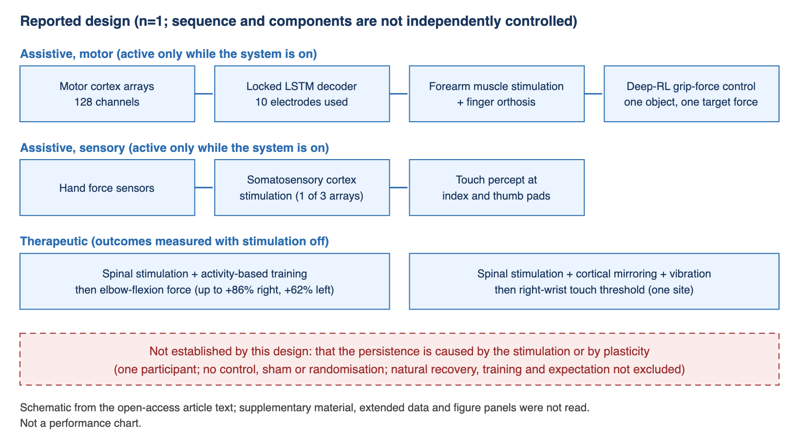
What was measured
Elbow force. After a baseline of about 20 weeks of weekly force recordings, which the authors describe as stable, tSCS paired with activity-based training was started. Median isometric elbow-flexion force rose from 13.25 N to 24.65 N on the right and from 24.03 N to 38.90 N on the left (medians over 10-week windows), which the paper reports as gains of up to 86% and 62% at about 35 weeks. The participant could then bring both hands to his face. These gains came before the cortical system was used for movement. Stimulation aimed at the hand and triceps produced no strength gain.
Grasping. A locked decoder, trained once and not retrained, was evaluated for about five months and reached accuracies of up to 84.6%, using 10 of 128 motor electrodes. With the reinforcement-learning agent on, grip force stayed under a set limit in 87% of trials versus 27% with it off (five sessions per condition), using hollow eggshells.
Touch. Light-touch detection at the right radial wrist improved during the tSCS plus cortical-mirroring period (monofilament thresholds down to about 10 g) and, per the authors, the gain persisted more than two months after stimulation ended. No sensation was reported at the index or thumb pads in any phase.
What the authors interpret
The authors write that the results “suggest” the intervention induced long-term plasticity changes in wrist sensitivity, and that electrode-specific neural changes “may have contributed”. These are interpretations of an uncontrolled single-participant sequence. We treat plasticity as a hypothesis, not a finding.
Deep dive
1. Background
The paper starts from two earlier lines of work. One is a motor neuroprosthesis in which an intracortical brain-computer interface (iBCI) is linked to real-time muscle stimulation to restore hand movement in complete tetraplegia; the paper cites earlier work from the same group (Bouton et al., Nature 2016) and another group (Ajiboye et al., Lancet 2017). The other is intracortical microstimulation (ICMS) of primary somatosensory cortex to give touch, which the authors note had mostly been used with robotic manipulators rather than the user’s own hand. The authors also state that temporary tactile enhancement has been shown but persistent recovery “has remained elusive”. They cite iBCI-mediated spinal stimulation restoring lower-limb function in incomplete injury (Lorach et al., Nature 2023). The study discussed here combines these elements, plus a reinforcement-learning layer, in one participant.
2. What this study newly reports
Participant and design. One man with a complete (ASIA Impairment Scale A) cervical injury, enrolled 13 months after injury, in a trial registered at ClinicalTrials.gov (NCT03680872) under an FDA Investigational Device Exemption and approved by the Northwell Health institutional review board. The study ran for more than three years, with experimental sessions one to three times per week. We omit further personal details.
Hardware. Five 10 x 10 platinum microelectrode arrays (Neuroport, Blackrock Neurotech) in the left hemisphere: two 64-channel arrays in motor cortex (128 channels) and three sparsely populated 32-channel arrays in S1 (96 channels). Only one S1 array evoked sensory percepts; the other two did not. Percepts were mostly at the index and thumb pads.
Stimulation parameters (as reported). S1 stimulation for grasp feedback: 100 µA, 300 µs pulse width, 100 µs interphase, 100 Hz for 1 to 2 s, on three electrodes. Cortical mirroring: 200 Hz, 5 s on and 5 s off, on up to 16 electrodes at once, paired with 120 Hz vibration for up to 10 minutes; the electrode set was updated every session. tSCS: a 10 kHz carrier delivered at 50 Hz, up to 300 mA, through a custom electrode patch targeted at C5-C6 or C7-C8 roots. The authors say benefits were seen with two sessions per week.
Decoder. Features were integrated broadband power (100 to 5,000 Hz) in 100 ms steps. Electrodes were selected for consistent modulation, leaving 10 of 128. The decoder was trained on data from a 16-day period and then locked. After locking, 39 three-class and 6 four-class data blocks were evaluated over about five months, with accuracies up to 84.6%. The decoder separates rest, open, close and (in four-class blocks) reach; the authors state that this is fewer degrees of freedom than other iBCI systems have shown and call it proof of principle. Note that the text gives the training period as six sessions in the Results and five in the Methods, so the exact count is unclear.
Grip force. The reinforcement-learning agent was trained in a simulation of the orthosis and hand and then used with the participant, who could see a plot of the grip force. Force stayed under the limit in 87% of trials with the agent on and 27% with it off (P = 0.012). With S1 feedback the participant reported object presence at 100% versus chance without feedback, in four feedback-on and three feedback-off blocks. The authors acknowledge the task used a single target force and a single type of object.
Sensation. Wrist assessments: 13 baseline, 11 intervention and 7 post-intervention monofilament sessions. Thresholds were better during the intervention than at baseline (P = 0.0011, Wilcoxon rank-sum, no multiple-comparison adjustment). In the post-intervention period the participant more often perceived a touch but mislocalised it (P = 0.0106 versus baseline), and intervention versus post-intervention did not differ (P = 0.29). In a blindfolded press-and-hold task at 700 g, wrist localisation averaged 81% over 11 sessions, unlike other sites. Before this phase, a 39-week tSCS-only regimen had not changed sensation.
Neural data. Modulation depth to touch rose more in stimulated than non-stimulated S1 electrodes (mixed-effects estimate 0.20, 95% CI 0.13 to 0.27, 960 electrode observations over 10 sessions), and the authors report a continued rise over 92 minutes after stimulation in an example dataset. Removing one outlier session lowered the estimate to 0.14 (95% CI 0.08 to 0.21) per the extended-data legend.
Anecdotal outcomes. The authors report self-reported daily-life gains outside sessions, which they describe as anecdotal.
Not reported in the text we read: an adverse-event summary or device-related safety outcomes; blinding of assessors; any sham or control condition. Supplementary material, extended data and figure panels were not read, so these may be addressed there.
3. Strengths and limitations of the method
Strengths.
- Objective outcome measures (load-cell force, monofilament thresholds, decoder accuracy per data block) rather than only self-report.
- A long pre-intervention force baseline (about 20 weeks, described as stable) and a longer follow-up than most first reports.
- Within-participant comparisons that give some internal checks: hand and triceps stimulation produced no strength gain; sensory change was confined to the wrist; one finger location (index volar pad) was left unstimulated as a control site; stimulated and non-stimulated electrodes were compared.
- Methods are described in detail, with a trial registration and device exemption number, and data and code are available on request.
- The authors state clear limits: single participant, single object and target force, and a “highly specialized system requiring a team of highly trained personnel to operate”.
Limitations.
- n=1. Results describe one person. The authors write that scaling will require addressing individual variability in stimulation mapping, decoder and sensory pattern selection.
- No control arm, sham or randomisation. The paper does not frame this as a limitation in the text we read. A stable pre-treatment baseline is useful, but the participant was about 13 months post-injury at enrolment and the study lasted more than three years, so continued natural recovery, practice and expectation remain possible explanations for gains that persist.
- Bundled interventions. Strength gains came from tSCS plus activity-based training; sensory gains from tSCS plus cortical mirroring plus vibration. The design cannot assign credit to any one component. Earlier phases preceded later ones, so order effects cannot be excluded (we have not seen the study timeline figure).
- The headline strength figures belong to the non-cortical component. The 86% and 62% are from tSCS with training, not from the brain-computer interface.
- Statistics. Tests treat repeated sessions within one person; the sensory comparisons were not adjusted for multiple testing; the wrist-sensitivity effect is at one site, and index and thumb sensation was not restored.
- Task scope. The eggshell task used a single target force; the decoder distinguishes three or four states; grasping relies on an orthosis because forearm muscle stimulation only partly activated the flexors and nerve-conduction results suggested lower motor neuron dysfunction.
- Internal text inconsistencies. The decoder training session count differs between sections, as noted above.
- Operational burden. The system needs trained staff; the authors suggest the reinforcement-learning and wearable tSCS parts could ease home use but do not show it.
4. Connections to neighbouring domains
Rehabilitation and neuromodulation. The paper’s own comparison point is non-invasive spinal stimulation for arm and hand function (for example Moritz et al., Nature Medicine 2024, as cited by the authors). The authors argue that a transcutaneous approach is easier to translate than epidural stimulation because of lower surgical burden. In this study the strength result came from the non-invasive part alone, which raises the question of what the implanted cortical component adds for therapy, as opposed to assistive use.
Machine learning. Two design choices stand out: a locked decoder that avoids retraining over about five months, and a small-state reinforcement-learning agent trained in simulation in a few episodes. Both are efforts to reduce maintenance and data demands, which are central problems for implanted interfaces. Evidence here covers one person, one object and a few decoder classes.
Plasticity research. The authors relate cortical mirroring to experiments on paired cortical stimulation and sensory input in animals and to biomimetic stimulation in humans. If activity-dependent plasticity does underlie the wrist result, it would link device neuromodulation to circuit-repair biology. This remains a hypothesis until a controlled comparison exists.
5. Commercialisation and investment perspective
TRL 4 (our estimate). A multi-component system was tested in a single participant in a clinical setting under a device exemption. Without more participants, controls or replication, we would not rate it higher. Individual parts (implanted arrays, microstimulation, transcutaneous stimulators) are more mature than the combined system.
Timeline (our estimate). An approved medical device would require multi-participant pivotal trials. Obstacles visible in the paper include the specialised staffing, the need to adapt mapping and decoding to each person, and unreported long-term implant data. We expect a decade or more, which is not a figure from the study.
Companies and funding. The study was funded by the New York State Department of Health Spinal Cord Injury Research Board (contract C37718GG) and the Feinstein Institutes for Medical Research at Northwell Health, with additional support from Blackrock Neurotech and Good Shepherd Rehabilitation Network. The arrays and stimulator used were Blackrock Neurotech products, and two Blackrock employees are thanked in the acknowledgements. The corresponding author reports financial interests in two companies (see Disclosure). We draw no investment inference; the paper is a feasibility report on one participant.
6. Counterarguments
- Causal reading of lasting recovery. That gains persisted after stimulation stopped does not show that stimulation produced them or that rewiring occurred. With one participant and no control or sham condition, natural recovery, training effects and expectation cannot be separated from stimulation effects. The authors’ own wording is hedged (“suggest”, “may have contributed”).
- What persisted. Strength gains are reported as lasting several months; the sensory gain is reported as lasting more than two months at one wrist site. After stimulation ended the participant more often mislocalised touch than correctly localised it, so the quality of the sensation is also open.
- Attribution to the cortical interface. The largest numbers come from non-invasive spinal stimulation with training. The cortical interface’s contribution is mainly assistive and measured while it is on.
- Generalisation. Injury level, completeness, age and time since injury could all change the response. Mapping and stimulation patterns were individually tuned.
- Safety and durability. We saw no adverse-event summary or electrode-longevity data in the text. Absence of a report is not evidence that none occurred.
- Source of interest. The corresponding author has financial interests and patents in the field (see Disclosure), and the developing institution funded the study. We attribute quantitative claims to the authors.
7. Indicators to watch
- Additional participants, with different injury profiles, and any control, sham or crossover design that could separate stimulation from natural recovery.
- Whether sensory gains extend beyond the wrist to the fingers, and whether localisation improves.
- A safety record: adverse events, electrode signal longevity and explant data.
- Tests of mechanism, such as imaging or electrophysiology with a control condition.
- Decoders with more degrees of freedom and multiple grip forces and objects.
- Home-use operation without trained staff, and the regulatory path for a multi-participant trial.
- Independent groups reporting comparable results.
References
The Nature Medicine article is open access; we read the article text. Supplementary material, extended data and figure panels were not read. The earlier works below are listed as cited in that article, with the citation details given there.
- Chandrasekaran, S., S. K. Wandelt, A. Jangam, et al. (25 authors). 2026. “A neuroprosthesis for restoring hand movement and sensation in a person with complete tetraplegia.” Nature Medicine 32: 2591-2601 (online 16 July 2026). DOI: 10.1038/s41591-026-04498-0; https://www.nature.com/articles/s41591-026-04498-0
- Preprint of the same study (medRxiv, not peer reviewed). 2025. “Restoring Cortically Mediated Movement and Sensation in Complete Tetraplegia.” DOI: 10.1101/2025.08.19.25330198
- Bouton, C. E., et al. 2016. “Restoring cortical control of functional movement in a human with quadriplegia.” Nature 533: 247-250. DOI: 10.1038/nature17435. (as cited in the Nature Medicine article; DOI matched in Crossref.)
- Ajiboye, A. B., et al. 2017. “Restoration of reaching and grasping movements through brain-controlled muscle stimulation in a person with tetraplegia: a proof-of-concept demonstration.” Lancet 389: 1821-1830. DOI: 10.1016/S0140-6736(17)30601-3. (as cited in the Nature Medicine article; DOI matched in Crossref.)
- Lorach, H., et al. 2023. “Walking naturally after spinal cord injury using a brain-spine interface.” Nature 618: 126-133. DOI: 10.1038/s41586-023-06094-5. (as cited in the Nature Medicine article; DOI matched in Crossref.)
- Moritz, C., et al. 2024. “Non-invasive spinal cord electrical stimulation for arm and hand function in chronic tetraplegia: a safety and efficacy trial.” Nature Medicine 30: 1276-1283. DOI: 10.1038/s41591-024-02940-9. (as cited in the Nature Medicine article; DOI matched in Crossref.)
- Press materials from Nature Portfolio, the Feinstein Institutes and trade media were consulted early on; figures in this post are taken from the article text instead, and where press figures differed from the article we used the article.
Disclosure
- This article is for informational purposes only and is not investment advice.
- The author holds no position in any company named in this post.
- Conflict-of-interest notes (from the article):
- The competing-interests statement says that Chad E. Bouton, the corresponding author, has financial interests in Neuvotion, Inc., a medical device company developing neurotechnologies for restoring function after stroke or spinal cord injury, and in Sanguistat, LLC, a company developing neurostimulation technologies to reduce blood loss. It also says he has multiple patents in the field of neuroprosthetics and related fields. The other authors declare no competing interests.
- Most authors are affiliated with the Feinstein Institutes for Medical Research, Northwell Health, which also funded the study (with the New York State Department of Health Spinal Cord Injury Research Board). The article lists additional support from Blackrock Neurotech and Good Shepherd Rehabilitation Network; Blackrock Neurotech made the arrays and the stimulator used. Other author affiliations include Baylor College of Medicine, Washington University, Johns Hopkins institutions and Lenox Hill Hospital.
- Quantitative results in this post are the authors’ reported results from one participant.
- Our reading covered the open-access article text; supplementary material, extended data and figure panels were not read.
- The figure in this article is an original drawing; no figure from the source was reproduced.


Leave a Reply